Table of Contents >> Show >> Hide
- What’s the difference between a 2D mammogram and a 3D mammogram?
- What the study found: why people are talking about 3D now
- Why 3D mammography can find more cancers (especially in dense breasts)
- Does 3D mammography save more lives?
- Tradeoffs: what 3D mammograms don’t magically fix
- Who benefits most from 3D mammography?
- Screening guidelines: when should you start, and how often?
- What to expect at a 3D mammogram appointment
- If something looks abnormal: what happens next?
- Bottom line: is a 3D mammogram “better” than a 2D scan?
- Real-world experiences with 3D mammograms (what people often notice)
If mammograms had a résumé, “find sneaky stuff early” would be listed in bold at the top. But here’s the twist:
traditional 2D mammography is basically a flat photo of a complicated, layered body part. And when
tissue overlaps (especially in dense breasts), important details can hide in plain sightlike trying
to spot a black cat in a dark closet… while wearing sunglasses.
A growing body of researchand a headline-making long-term studysuggests that 3D mammograms
(also called digital breast tomosynthesis or DBT) can detect more cancers and reduce
“call-backs” compared with standard 2D scans. That’s a big deal, because fewer call-backs can mean less anxiety,
fewer extra appointments, and fewer “so, about that mammogram…” phone calls during lunch.
Let’s break down what 3D mammography is, what the research actually shows, who benefits most, and what tradeoffs
(cost, radiation, follow-up testing) are part of the full storywithout turning your brain into medical oatmeal.
What’s the difference between a 2D mammogram and a 3D mammogram?
2D mammography: the classic “two-view photo”
A standard 2D mammogram takes X-ray images of each breast from two angles (think “top-to-bottom” and “side-to-side”).
It’s fast, widely available, and has helped reduce breast cancer deaths through early detection. But because it’s
a flat image, overlapping tissue can create shadows or hide small abnormalitiesespecially if breast tissue is dense.
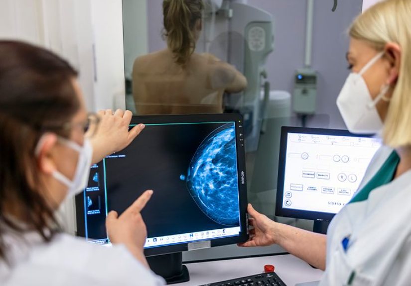
3D mammography (DBT): the “flip-book” view of your breast tissue
A 3D mammogram still uses X-rays and the same brief compression, but the machine takes multiple images from different
angles. A computer reconstructs them into thin “slices,” giving radiologists a layered view of the breast.
In practice, this makes it easier to see around overlapping tissue and better evaluate subtle distortions or small masses.
In other words: 2D is a single snapshot. 3D is more like scrolling through layersless “Where’s Waldo?” and more
“Oh, there you are.”
What the study found: why people are talking about 3D now
The reason 3D mammography keeps showing up in the news is simple: outcomes that matter to real humans are improving.
In a large long-term analysis of screening data (covering years of real-world mammograms), researchers reported that
screening with DBT was linked to:
- Higher cancer detection (more cancers found during screening)
- Lower recall rates (fewer people called back for extra imaging that turns out to be benign)
- Fewer advanced cancers at diagnosis (suggesting some cancers are being found earlier)
One widely cited long-term study described a “win, win, win” pattern: DBT had a higher cancer detection rate
(often described around 5.3 per 1,000 screens versus about 4.0 per 1,000 with 2D in that dataset),
a lower recall rate (about 7.2% versus 10.6%), and a lower proportion of advanced cancers.
Translation: more true problems found, fewer false alarms, and potentially earlier-stage detection.
That doesn’t mean 2D is “bad.” It means 3D may do a better job in common real-life conditionsespecially when tissue overlap
makes the 2D image harder to interpret.
Why 3D mammography can find more cancers (especially in dense breasts)
Dense tissue can “camouflage” tumors on 2D images
Dense breast tissue contains more fibrous and glandular tissue than fat. On a mammogram, dense tissue looks white.
Unfortunately, many tumors also look white. That “same-color problem” can make small cancers harder to see on 2D mammograms.
3D mammography helps by reducing the visual clutter of overlapping tissue. When radiologists can scroll through thin slices,
suspicious areas are less likely to be hidden behind dense tissue patterns.
DBT is often better at showing masses and distortions
Research and clinical experience suggest DBT is particularly good at clarifying masses and
architectural distortions (subtle “pulling” patterns in tissue). Some cancers that are hard to notice
on a flat image become more obvious in the layered 3D view.
DBT doesn’t automatically “solve” every challenge. Tiny calcifications can still be visible on both 2D and 3D,
and sometimes additional views are needed either way. But overall, the 3D approach improves clarity in many cases.
Does 3D mammography save more lives?
Here’s where we stay honest: while DBT is better at finding tumors and reduces call-backs in many studies,
it’s still being studied whether DBT leads to a clear additional reduction in breast cancer deaths compared with standard mammography.
Big, carefully designed trials are underway to answer that question more definitively. Meanwhile, major health organizations generally
recognize that both 2D and 3D mammography are effective screening optionsand that access, follow-up, and consistent screening matter a lot.
Tradeoffs: what 3D mammograms don’t magically fix
1) False positives still exist (just often fewer of them)
Any screening test can flag something that turns out to be benign. DBT often lowers recall rates, but it doesn’t eliminate
false positives. Some people will still need additional imaging (extra mammogram views and/or ultrasound), and a smaller number
will need a biopsy to confirm what’s going on.
2) Overdiagnosis is a real discussion
Overdiagnosis means finding a cancer that would not have caused harm during a person’s lifetime. This is a complicated topic and
one reason screening guidelines sometimes differ. The encouraging news from long-term DBT research is that some datasets show DBT
may be finding “bad actors” earlier rather than simply finding more low-risk diseasebut the broader question is still being studied.
3) Radiation dose: usually small, but worth understanding
Mammography uses low-dose radiation. Some 3D protocols can slightly increase exposure if both separate 2D and 3D images are taken.
Many centers use “synthetic 2D” images generated from the 3D data to keep dose closer to standard screening levels.
If you’re concerned, ask the imaging center what technique they use.
4) Cost and coverage vary
3D mammograms can cost more than 2D, and coverage can vary by insurer and plan. In many places, DBT is widely covered, but “widely”
isn’t the same as “always,” so it’s smart to confirm coverage before your appointment if cost is a concern.
Who benefits most from 3D mammography?
DBT can be helpful for many people, but the added value is often greatest when tissue overlap is most likely to confuse the picture.
Common situations where 3D may be especially useful include:
- Dense breasts, where small tumors can be harder to see on 2D images
- Prior call-backs for overlapping tissue or benign findings (DBT may reduce repeat “false alarm” cycles)
- Higher-than-average risk (family history, prior high-risk biopsy results, etc.), as part of a personalized plan
- When the facility has strong DBT experience (technology plus expert interpretation is the real power combo)
If you’re at high risk (for example, a strong family history or a known genetic mutation), screening may involve more than mammography.
Some guidelines recommend adding breast MRI to annual mammograms for certain high-risk groups. This is where a risk conversation
with your clinician really matters.
Screening guidelines: when should you start, and how often?
You may see different recommendations depending on the organization, the person’s risk, and how harms (like false positives) are weighed.
Here’s the practical takeaway: pick a credible guideline, then personalize it with your risk factors.
Average risk
- Some major U.S. recommendations support biennial screening from ages 40–74 for average-risk individuals.
- Other organizations support annual screening starting at 40 (with the option to adjust frequency later).
If you’re thinking, “Cool, so which is it?”you’re not alone. The best choice often depends on personal values (tolerance for false positives),
access to follow-up care, and individual risk. The most important thing is not to let confusion delay screening for years.
Higher-than-average risk
People with higher risk may start earlier and/or use additional imaging (like MRI). Risk is not just “family history yes/no.”
It can involve:
- Genetic mutations (such as BRCA-related mutations)
- History of chest radiation at a young age
- Strong family history patterns
- Prior biopsy results showing certain high-risk lesions
What to expect at a 3D mammogram appointment
The appointment is similar to a 2D mammogram. Expect brief compression (unpleasant, not dangerous), and the imaging takes seconds per view.
Many people say the anticipation is worse than the testkind of like the dentist chair, but with fewer tiny hooks and more awkward angles.
Quick prep tips
- Avoid deodorant, powders, or lotions on your underarms/chest that day (they can show up on images).
- If your breasts are tender, scheduling when they’re less sensitive may help.
- Bring prior imaging info if you’ve switched facilitiescomparisons matter.
Your report may mention breast density
Breast density is commonly categorized into four levels (from mostly fatty to extremely dense). Many U.S. facilities now provide
standardized breast density notifications in patient summaries. If you’re told you have dense breasts, it does not mean you have cancer.
It means mammography can be less sensitive and your risk profile may be differentworth a conversation, not a panic spiral.
If something looks abnormal: what happens next?
A screening mammogram (2D or 3D) is a first look. If the radiologist sees something unclear or suspicious, you may be asked back for:
- Diagnostic mammogram views (more targeted images)
- Ultrasound to better characterize a mass
- MRI in select situations, especially for higher-risk individuals
- Biopsy if imaging suggests it’s needed to confirm the diagnosis
Most call-backs do not end in a cancer diagnosis. Still, follow-up mattersbecause the whole point of screening is to catch serious problems early,
when treatment can be simpler and outcomes are better.
Bottom line: is a 3D mammogram “better” than a 2D scan?
For many people, yes3D mammography improves cancer detection and reduces call-backs compared with 2D mammography in multiple studies,
including long-term real-world data. It appears especially helpful when breast density or overlapping tissue makes interpretation difficult.
The fairest summary is this: 3D mammography is a meaningful upgrade for screening accuracy and the patient experience (fewer “false alarm” call-backs),
but it’s not a magic shield. It still requires follow-up systems, equitable access, and personalized decision-makingespecially for people with dense breasts or higher risk.
If you’re choosing between 2D and 3D, a good question is: “What does my risk look like, what does my facility offer well,
and what will my insurance cover?” Then you make the call with real informationnot just vibes and fear.
Real-world experiences with 3D mammograms (what people often notice)
Research statistics are helpful, but real life is messy. People don’t experience “a 3.4% recall reduction”they experience
a phone call, a calendar scramble, and a stomach drop that could qualify as an Olympic sport. That’s why one of the most
meaningful “everyday” differences with 3D mammography is how it can change the emotional rhythm of screening.
Fewer call-backs can feel like getting your week back. Many patients who’ve had multiple screening cycles describe
call-backs as the most stressful partnot because additional imaging is painful, but because waiting is. Waiting for the follow-up appointment,
waiting for the radiologist’s review, waiting for the final “all clear.” When 3D mammography reduces the number of false alarms from tissue overlap,
people often describe it as a quieter screening experience: fewer unexpected appointments, fewer time-off requests, fewer “I guess I’ll just
refresh my inbox every 9 minutes” moments.
Dense breast notifications can be confusing at first. A common story goes like this: someone opens a mammogram result that says
“heterogeneously dense” or “extremely dense,” and suddenly it feels like their own body just added a surprise plot twist.
Many people report that the first question is, “Is this dangerous?” The second is, “Why didn’t anyone tell me sooner?”
In reality, density is common, can change over time, and can’t be “felt” during self-exams. What it does change is how clearly a mammogram
can see through tissueand that’s where 3D mammography often becomes part of a practical next step.
Some patients describe 3D as more reassuringothers don’t notice a difference. The physical experience is usually similar:
brief compression, a few images, done. Some people say the 3D exam feels slightly longer because the machine is acquiring more angles.
But the most noticeable differences tend to show up after the appointment: clearer results, fewer “we need one more view,” fewer repeats due to overlap.
For others, the experience feels identicaland that’s also a valid outcome if it still delivers a solid screening.
Clinicians often talk about clarity and confidence. Radiologists and technologists commonly describe 3D mammography as a tool that reduces
uncertainty in busy screening settings. Instead of debating whether a shadow is real or just overlapping tissue, they can scroll through slices and see
whether something truly persists across layers. When something is real, 3D can make margins and shapes easier to evaluatehelping triage who needs diagnostic
imaging and who can safely go back to normal screening.
Insurance and access can be the un-funniest part of the story. People sometimes assume that “newer and better” automatically means “covered.”
In many areas it is, but coverage can still vary by plan and region. A common experience is calling the imaging center to confirm: Is 3D available? Is it standard?
Is there a separate charge? This step isn’t glamorous, but it can prevent surprise bills. Patients who’ve been through it often recommend asking directly,
“Will I have out-of-pocket costs for DBT?” and getting the answer in writing if possible.
Ultimately, the most consistent “experience takeaway” is this: screening works best when it’s regular, accessible, and followed up appropriately.
3D mammography can improve detection and reduce call-backs, but the real win is when the system around itclear communication, timely diagnostic follow-up,
and personalized risk planningworks smoothly enough that people can get screened, get answers, and get on with their lives.