Table of Contents >> Show >> Hide
- First, what do “white spots” actually mean?
- Common causes of white spots on skin
- 1) Tinea versicolor (a yeast overgrowth)
- 2) Vitiligo (autoimmune loss of pigment)
- 3) Pityriasis alba (often linked with eczema in kids and teens)
- 4) Post-inflammatory hypopigmentation (the “after party” of skin inflammation)
- 5) Idiopathic guttate hypomelanosis (tiny “white sunspots”)
- 6) Less common (but important) possibilities
- How dermatologists figure out the cause
- Treatments that match the cause (what actually helps)
- When to see a clinician (a quick “don’t ignore this” list)
- Everyday care that helps no matter the diagnosis
- Conclusion
- Experiences people commonly report (and what they often learn)
You glance down at your arm andsurprisethere’s a pale patch that wasn’t invited to the party. “Manchas blancas en la piel” literally means
white spots on the skin, and it’s a common concern in the U.S. dermatology world. The tricky part: “white spots” is a description, not a diagnosis.
Sometimes it’s a harmless sun-related change. Other times it’s a yeast overgrowth, leftover pigment changes after a rash, or (yes) a condition like vitiligo.
This guide breaks down the most common causes of white patches and light spots, how clinicians tell them apart, and what treatments actually help.
Expect practical tips, clear explanations, and exactly zero fearmongering. (Your skin deserves better.)
Medical note: This article is educational and not a substitute for medical care. If a patch is spreading fast, painful, bleeding, or shows up with other symptoms, get checked by a clinician.
First, what do “white spots” actually mean?
Most “white spots” fall into one of two categories:
- Hypopigmentation: the skin has less pigment than the surrounding area (lighter, but not completely milk-white).
- Depigmentation: pigment is largely gone (often brighter white and sharper-edged), as seen in many cases of vitiligo.
The difference matters because it changes what causes are most likelyand what treatments are worth your time and money.
Also, lighting can lie. A bathroom bulb can make anything look suspicious, including your life choices.
Common causes of white spots on skin
Below are the heavy hitters clinicians consider in the U.S. (Translation: the usual suspects.)
1) Tinea versicolor (a yeast overgrowth)
Despite the name, this isn’t a “worm” and it isn’t your hygiene. Tinea versicolor happens when a yeast that normally lives on skin grows too much.
It can cause patches that look lighter (or sometimes darker) than your normal tone, often on the chest, back, shoulders, and neck. A clue is
fine, powdery scaleespecially if you gently scratch and see a subtle “dusting.”
It’s more common in warm, humid conditions, in people who sweat a lot, and during summer months. The fun twist: even after the yeast is treated,
the lighter color can linger for weeks because pigment takes time to normalize.
Typical treatment: topical antifungals (shampoos/washes/creams) such as selenium sulfide, ketoconazole, or zinc pyrithione. For widespread or stubborn cases, a clinician may consider oral antifungals.
2) Vitiligo (autoimmune loss of pigment)
Vitiligo causes the immune system to target melanocytes (the cells that make pigment), leading to well-defined patches that can slowly expand over time.
It can affect any area, including hands, face, body folds, and hair (hair can turn white in affected areas). Vitiligo is not contagious
and not dangerous in a “this will harm your organs tomorrow” way, but it can be emotionally heavyand sun sensitivity is a real concern because pigment helps protect against UV.
There’s no single “cure,” but there are meaningful options to restore pigment in some people, slow progression, and reduce contrast. Treatment choice often depends on where the patches are, how long they’ve been present, and how active the condition is.
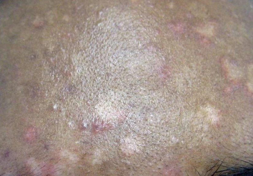
3) Pityriasis alba (often linked with eczema in kids and teens)
Pityriasis alba commonly shows up as lighter, slightly scaly patchesoften on the face, arms, or upper bodyespecially in children and adolescents.
It’s frequently connected to eczema or dry skin inflammation. When surrounding skin tans, the lighter patches can look more dramatic, which is rude but predictable.
The good news: it’s harmless and often improves over time. The goal is usually to calm dryness and inflammation so pigment can recover naturally.
4) Post-inflammatory hypopigmentation (the “after party” of skin inflammation)
Any rash, injury, burn, procedure, or inflammation (eczema, psoriasis, acne, infections) can temporarily disrupt pigment production.
The result can be lighter areas exactly where the skin was irritated before. Many cases gradually improve over months once the underlying issue is controlled
especially when you protect the area from the sun.
5) Idiopathic guttate hypomelanosis (tiny “white sunspots”)
These are small (often a few millimeters), flat, pale-to-white dots that commonly appear on sun-exposed areas like shins and forearms, especially with age.
They’re benign and thought to be related to cumulative sun exposure and natural skin aging changes. Many people notice them most after spending time outdoors.
Treatment is usually optional. If you’re bothered cosmetically, a dermatologist can discuss options, but expectations should be realisticthis is more “manage” than “erase.”
6) Less common (but important) possibilities
- Medication or steroid-related lightening: strong topical steroids or injections can sometimes cause localized light spots.
- Contact/chemical leukoderma: repeated exposure to certain chemicals can disrupt pigment in localized areas.
- Other skin diseases: psoriasis, eczema variants, and certain inflammatory conditions can leave lighter patches as they heal.
- Rare conditions: congenital pigment differences (present from childhood) or genetic pigment disorders.
How dermatologists figure out the cause
If you’ve ever wished your skin came with a user manual, you’re not alone. Clinicians use a mix of pattern recognition and a few simple tools:
- History: When did it start? Is it itchy? Any new products, sweating, recent rash, sunburn, or skin injury?
- Visual exam: Borders (sharp vs. blurry), location (trunk vs. face vs. hands), scale, and symmetry.
- Wood’s lamp: A special light that can make certain pigment patterns and fungal changes more noticeable.
- Skin scraping (KOH test): A quick microscope test for fungal/yeast causes like tinea versicolor.
- Biopsy (sometimes): If the diagnosis isn’t clear, a small sample can confirm what’s happening in the pigment layer.
Translation: you usually don’t need a thousand lab tests. But you do need the right diagnosisbecause “random cream roulette” is expensive and emotionally exhausting.
Treatments that match the cause (what actually helps)
Tinea versicolor: antifungals + patience
- Topical antifungal washes (often first-line): selenium sulfide, ketoconazole, or zinc pyrithione as directed.
- Topical antifungal creams: options like clotrimazole or other antifungals depending on severity.
- Oral antifungals: sometimes used for widespread or recurrent cases under medical supervision.
A common frustration: the yeast may be gone, but the light spots remain temporarily. That doesn’t always mean treatment failedpigment just needs time to catch up.
Vitiligo: repigmentation options + skin protection
Vitiligo treatment is typically individualized. Options may include:
- Topical corticosteroids (often for newer or localized patches): can help restore pigment in some cases.
- Topical calcineurin inhibitors (like tacrolimus/pimecrolimus): often used on sensitive areas (e.g., face) depending on clinician guidance.
- Light therapy (such as narrowband UVB phototherapy): used to stimulate repigmentation; typically requires repeated sessions.
- Topical ruxolitinib cream (a JAK inhibitor): FDA-approved for nonsegmental vitiligo in patients aged 12 and older; results may take months and must be used exactly as prescribed.
- Camouflage: makeup or self-tanners can reduce contrast immediately (and your schedule doesn’t have to revolve around phototherapy appointments).
Sun protection is non-negotiable: depigmented areas burn more easily, and sunburn can worsen contrast. Broad-spectrum sunscreen, protective clothing, and shade are your skin’s best friends.
Pityriasis alba: moisturize, calm inflammation, avoid over-treating
- Moisturizers: basic, consistent emollients improve dryness and reduce visible scale.
- Anti-inflammatory topicals: mild topical steroids or non-steroid anti-inflammatories may be used if itchy or inflamed (especially when eczema is active).
- Sun protection: helps reduce contrast between surrounding tanned skin and lighter patches.
The goal isn’t “bleach the whole face to match.” The goal is restoring the skin barrier so pigment can normalize.
Post-inflammatory hypopigmentation: treat the trigger, then protect
The most effective strategy is usually boring (which is secretly great): identify and control the underlying inflammation, then give pigment time to recover.
Clinicians may use anti-inflammatory topicals when appropriate and emphasize sun protection to prevent the surrounding skin from darkening and making contrast worse.
Idiopathic guttate hypomelanosis: manage expectations
These small white sunspots are benign and often don’t require treatment. If you want to address them cosmetically, a dermatologist can discuss options.
What matters most is sun protectionboth for appearance and overall skin health.
When to see a clinician (a quick “don’t ignore this” list)
Make an appointment sooner rather than later if:
- The patch is spreading quickly or new patches are appearing rapidly.
- You have white patches on the face, hands, genitals, or around body folds and you’re unsure of the cause.
- There’s pain, bleeding, crusting, significant itch, or signs of infection.
- You have other autoimmune conditions (or a strong family history) and notice sharply defined white areas.
- A child develops new or changing patches and you want clarity (and peace of mind).
Everyday care that helps no matter the diagnosis
- Use broad-spectrum sunscreen daily on exposed areas. It reduces sunburn risk and helps prevent contrast from getting worse.
- Be gentle: harsh scrubs and aggressive “brightening” routines can trigger irritation and pigment changes.
- Moisturize consistently, especially if there’s dryness or eczema.
- Don’t self-prescribe strong steroids for long periodsincorrect use can thin skin and sometimes cause localized lightening.
- Track changes: take photos monthly in consistent lighting. This helps you (and your dermatologist) see trends without guessing.
Conclusion
White spots on the skin can be anything from a common yeast issue (tinea versicolor) to inflammation-related pigment changes, to vitiligo.
The most useful next step isn’t panicit’s pattern recognition and, when needed, a quick clinical exam. Once you know the cause, treatments are usually straightforward:
antifungals for tinea versicolor, barrier repair and anti-inflammatories for pityriasis alba, time and trigger control for post-inflammatory hypopigmentation,
and targeted options (including topical medications and light therapy) for vitiligo.
If the spots are spreading, sharply defined, or stressing you out, that’s reason enough to get help. Skin changes are commonsuffering in silence is optional.
Experiences people commonly report (and what they often learn)
People usually don’t wake up thinking, “Today seems like a great day for mysterious white patches.” The first experience many report is the
double-take moment: you catch a glimpse in bright daylight, realize something looks lighter, then immediately test every lighting angle in your house like
you’re filming a detective show. That reaction is normalskin is personal, and changes can feel louder than they are.
One common story: someone notices light patches on the upper back after a hot summer of workouts. They assume it’s “sun damage,” but the patches have a subtle
scale and don’t tan like the surrounding skin. A clinician confirms tinea versicolor, and an antifungal wash clears the yeast.
The surprising part? The color doesn’t snap back overnight. Many people report a few weeks of “Is it still there?” anxiety, even though the infection is controlled.
Learning that pigment recovery can lag behind treatment is often a huge relief (and saves people from over-treating their skin out of frustration).
Another frequent experience shows up in families: a parent sees pale patches on a child’s cheeks or arms, especially after vacation.
The child isn’t sick, but the patches look more obvious next to a tan. This often ends up being pityriasis albadryness/eczema-related inflammation.
Parents commonly say the most helpful advice wasn’t a “miracle cream,” but a routine: gentle cleanser, consistent moisturizer, and sunscreen.
Once they stop scrubbing the area like it owes them money, the skin barrier settles down and the contrast fades over time.
Then there’s the “leftover mark” experience. Someone has a rash, a burn, acne flare-ups, or an eczema season that finally improves… and then light spots remain.
That’s classic post-inflammatory hypopigmentation. People often describe it as unfair: “I did everything rightwhy do I still have a reminder?”
What helps is understanding that pigment cells can be temporarily sluggish after inflammation. Many clinicians emphasize treating the underlying trigger first,
then protecting the area from sun so the surrounding skin doesn’t darken and make the contrast worse. With time, many of these patches gradually blend back in.
Vitiligo experiences can feel different because the contrast may be sharper and the emotional impact heavier. People commonly report noticing a small patch
near the hands, eyes, or mouth that slowly becomes more obvious. Some feel fine at first, then get hit with it laterlike a delayed reaction.
Others want to act quickly. A practical takeaway many people share is that a supportive dermatologist makes a big difference:
someone who discusses options (topicals, light therapy, newer prescriptions when appropriate), sets realistic expectations, and doesn’t treat you like you’re “overreacting.”
Even for people who choose not to pursue repigmentation, learning sun-protection habits and finding camouflage options (if desired) can restore a sense of control.
Across all these experiences, the pattern is consistent: the most stressful phase is often uncertainty. Once people know the likely cause and the plan,
the anxiety usually drops. The goal isn’t perfect skinno one has that. The goal is clear answers, safer routines, and treatments that match what your skin is actually doing.