Table of Contents >> Show >> Hide
- What is paraseptal emphysema?
- How it differs from other emphysema types (and why you should care)
- Causes and risk factors
- Symptoms: what you might notice (and what you might not)
- Complications and related conditions
- How doctors diagnose paraseptal emphysema
- Treatment and management
- Outlook: what to expect over time
- Living well with paraseptal emphysema
- When to see a doctor
- FAQ
- Experiences related to paraseptal emphysema (what people commonly report)
- SEO Tags
If lungs were sponges, emphysema is what happens when the sponge’s tiny holes stretch out, merge together, and lose their spring.
Paraseptal emphysema is a specific pattern of that damageone that likes to hang out near the outer edge of the lung.
It can be quiet for years… until it isn’t. (Yes, your lungs can be drama queens.)
This guide breaks down what paraseptal emphysema is, what symptoms to watch for, what doctors look for on imaging and breathing tests,
and what “outlook” really means in everyday life. It’s educational, not a diagnosisif you’re worried about symptoms, a clinician is the right next stop.
What is paraseptal emphysema?
Paraseptal emphysema (also called distal acinar emphysema) is a subtype of emphysema where the airspace enlargement
happens mainly at the edges of the lung, close to the pleura (the lining around the lung) and along connective tissue “septa” that divide lung regions.
Think: “perimeter problem,” not “center of the lung” problem. [1]
Because the changes are often localized and peripheral, people can have it without obvious day-to-day symptomsespecially early on. [1]
What makes it clinically important is that it may form or sit next to thin-walled air pockets called blebs or larger ones called bullae,
which can sometimes rupture and cause a spontaneous pneumothorax (collapsed lung). [1][9]
Where in the lungs does it show up?
Paraseptal emphysema is commonly seen near the lung surface and can be more noticeable in upper lung regions on CT imaging. [1]
It can occur by itself, but it may also coexist with other emphysema patterns (like centrilobular emphysema) in people who have broader COPD changes. [1]
How it differs from other emphysema types (and why you should care)
Emphysema isn’t one-size-fits-all. The “type” helps clinicians describe where the damage is most prominent and anticipate certain risks:
- Centrilobular emphysema: more central in the lung lobule; commonly linked to smoking and classic COPD airflow limitation.
- Panacinar (panlobular) emphysema: more uniform involvement; can be associated with alpha-1 antitrypsin deficiency.
- Paraseptal (distal acinar) emphysema: peripheral involvement; often discussed in relation to blebs/bullae and pneumothorax risk. [1][9]
Why it matters: two people can both be told they “have emphysema,” but their symptoms, complications, and management priorities may differ.
Paraseptal emphysema especially raises the question, “Is there a risk of blebs/bullae and sudden collapse?” [9]
Causes and risk factors
Emphysema is strongly associated with exposures that irritate and inflame the lungs over timemost famously, tobacco smoke.
But not everyone with emphysema has a smoking history, and risk can stack from multiple directions. [3]
Common risk drivers
- Smoking (current or past): the most common cause of emphysema overall. [4]
- Secondhand smoke and early-life exposure: can affect lung development and long-term risk. [3]
- Air pollutants and workplace irritants: chemical fumes, dust, and other toxins can contribute. [4]
- Genetic factors: alpha-1 antitrypsin deficiency is a known inherited risk for emphysema, often prompting screening in certain cases. [8]
What about vaping or marijuana?
Research and clinical reporting continue to evolve. Some clinical resources note that e-cigarettes introduce inhaled chemicals that may contribute to lung injury and may be discussed as a potential contributor in emphysema-related education. [7]
Marijuana smoke exposure is also discussed in the context of blebs/bullae and spontaneous pneumothorax in some medical references. [9]
Bottom line: if it’s smoke or aerosolized chemicals going into your lungs, your lungs are not sending a thank-you card.
Symptoms: what you might notice (and what you might not)
Paraseptal emphysema can be asymptomatic for a long time, especially when limited in extent. [1]
When symptoms show up, they often resemble emphysema/COPD symptoms in generalparticularly if other COPD changes are present. [4][8]
Possible symptoms
- Shortness of breath (often worse with activity) [8]
- Chronic cough (sometimes with mucus) [8]
- Wheezing [8]
- Fatigue and reduced exercise tolerance [8]
- Unintended weight loss or sleep issues in more advanced disease [8]
Red-flag symptoms: possible pneumothorax
Because paraseptal emphysema can be associated with blebs/bullae near the pleura, a rupture can let air into the pleural space and collapse part of the lung. [9]
Seek urgent medical care if you have:
- Sudden shortness of breath
- Sharp, pleuritic chest pain (pain that worsens with breathing)
- Feeling faint, severe distress, or rapidly worsening symptoms
Complications and related conditions
The biggest “headline complication” discussed with paraseptal emphysema is spontaneous pneumothoraxespecially in younger adults when it occurs independently. [1]
But there are other practical issues that can come along for the ride, especially if COPD is present.
Possible complications
- Spontaneous pneumothorax (collapsed lung) [1][9]
- Bullous disease (large bullae that can reduce effective breathing space) [10]
- Progressive airflow limitation if emphysema is part of COPD [11]
- Lower oxygen levels in more advanced disease (sometimes requiring oxygen therapy) [6]
How doctors diagnose paraseptal emphysema
A clinician usually combines symptoms, risk history (like smoking or occupational exposure), a physical exam, breathing tests, and imaging.
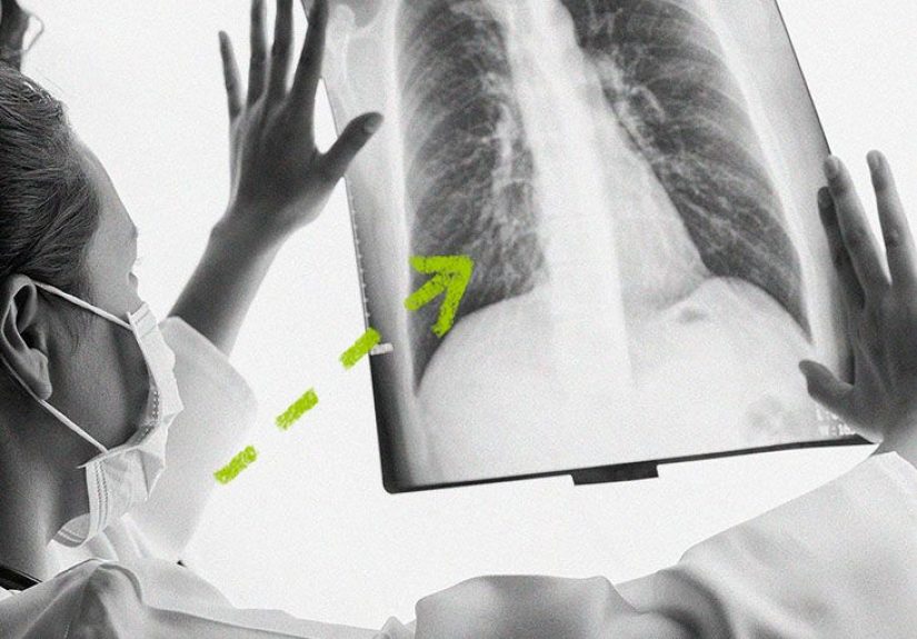
Importantly, paraseptal emphysema is often best characterized on a CT scan. [6][8]
Common tests
- Spirometry / pulmonary function tests: measure airflow limitation and help grade severity. [6][8]
- Chest X-ray: may show hyperinflation or other changes, but can miss subtle emphysema patterns. [6]
- CT scan: provides detail on emphysema distribution, blebs/bullae, and other lung findings. [8]
- Pulse oximetry / arterial blood gas: checks oxygenation when needed. [6]
- Alpha-1 antitrypsin deficiency screening: considered in select patients (especially early onset or strong family pattern). [8]
A realistic example
Someone gets a CT scan for an unrelated reasonsay, a persistent cough, a lung nodule follow-up, or even a pre-surgery evaluation.
The radiology report notes “subpleural emphysematous change” or “paraseptal emphysema” near the lung apices. The person feels mostly fine,
but the clinician uses that finding as a reason to ask deeper questions about exposures, breathing symptoms, and prevention steps.
Treatment and management
There’s no single “cure” that reverses emphysema, but there’s a lot that can be done to slow progression, reduce symptoms, and lower complication risk.
Management is individualizedbased on symptoms, lung function, oxygen levels, and whether bullae/pneumothorax risk is present. [6][7]
1) The cornerstone: stop lung irritation
- Quit smoking (if you smoke). This is the single biggest lever for slowing COPD/emphysema progression. [3][4]
- Avoid secondhand smoke and workplace irritants when possible. [3]
- Get vaccines (flu and pneumococcal are commonly recommended for people at risk of serious respiratory complications). [7]
2) Medications (when needed)
If symptoms or airflow limitation are present, clinicians may use inhaled medications commonly used in COPD care:
bronchodilators (to open airways) and sometimes inhaled steroids in specific situations. [7]
Antibiotics may be used for bacterial infections that worsen symptoms, when appropriate. [7]
3) Pulmonary rehabilitation
Pulmonary rehab is a structured program combining education, exercise training, breathing techniques, and supportoften helping people do more with less breathlessness. [5][6]
It’s one of those underrated “this actually changes daily life” interventions.
4) Oxygen therapy (for low oxygen levels)
If blood oxygen is low, supplemental oxygen can improve symptoms and quality of lifeand for some people with severe disease, it can improve outcomes.
The plan may be “only during activity,” “only during sleep,” or “most of the time,” depending on testing. [6]
5) Procedures and surgery (selected cases)
When large bullae interfere with breathing, clinicians may consider procedures such as removal of bullae (bullectomy), or other specialized interventions in carefully selected patients. [7][10]
Some emphysema patients may be evaluated for lung volume reduction surgery, certain bronchoscopic approaches, or (rarely) transplant when disease is very advanced. [6][7]
Outlook: what to expect over time
“Outlook” depends on a few key variables:
how extensive the emphysema is, whether COPD airflow limitation is present, smoking/exposure status, oxygen levels, activity tolerance, and other health conditions.
Some people with limited paraseptal emphysema remain stable and minimally symptomatic for years. [1]
Factors linked with a better outlook
- Stopping smoking and avoiding irritants [3]
- Early evaluation and treating symptoms before deconditioning sets in
- Consistent pulmonary rehab and activity [5]
- Up-to-date vaccines to reduce severe respiratory infections [7]
What about pneumothorax risk?
If you have blebs/bullae and a history of pneumothorax, your clinician may give specific guidance about recurrence prevention, symptom monitoring,
and activities that may change pressure dynamics (for example, certain high-altitude situations or diving can be relevant to pneumothorax discussions). [9]
This is one of those “personalized medicine” zonesyour imaging and history matter a lot.
Living well with paraseptal emphysema
Even when lung structure can’t be “un-damaged,” daily function can improvesometimes dramaticallywhen you build the right routine.
Here are practical, clinician-aligned habits that many care plans include:
Breathing and pacing
- Pursed-lip breathing during exertion to reduce air trapping
- Slow starts: give your lungs a warm-up before stairs or brisk walking
- Plan recovery time after big tasks (groceries, cleaning, long walks)
Fitness and nutrition
- Use pulmonary rehab principles: steady, supervised progression beats “weekend warrior” bursts. [5]
- Prioritize protein and balanced calories; unintended weight loss can be a problem in advanced disease. [8]
- Stay hydrated if mucus is an issue (ask your clinician if you have fluid restrictions).
Protect your lungs like they’re VIPs
- Avoid smoke exposure at home and in social settings. [3]
- Use appropriate workplace protection if you’re around dust/fumes.
- Have a plan for respiratory infections (when to call, when to test, what to do).
When to see a doctor
Make an appointment if you have persistent shortness of breath, chronic cough, wheezing, or reduced exercise toleranceespecially with a history of smoking or exposure risks. [4][8]
Seek urgent care for sudden chest pain and sudden shortness of breath (possible pneumothorax). [9]
FAQ
Is paraseptal emphysema the same as COPD?
Not exactly. Paraseptal emphysema is a pattern of emphysema. COPD is a broader diagnosis involving persistent airflow limitation, often with emphysema and/or chronic bronchitis.
Some people with paraseptal emphysema meet criteria for COPD, and others don’t. [4][11]
Can paraseptal emphysema be reversed?
Emphysema-related structural damage is generally considered permanent, but symptoms and function can improve with smoking cessation, medications (when needed),
pulmonary rehab, and oxygen therapy when indicated. [6][7]
If I feel fine, do I need to do anything?
If it’s an incidental CT finding, your clinician may still recommend prevention steps (especially avoiding smoke), baseline breathing tests,
and guidance on warning signs of pneumothorax. “Feeling fine” is greatthink of it as a head start.
Does everyone with paraseptal emphysema get a collapsed lung?
No. But because this pattern can be associated with peripheral blebs/bullae, clinicians pay attention to pneumothorax risk and symptoms that could signal one. [1][9]
Experiences related to paraseptal emphysema (what people commonly report)
The experiences below are not medical advice and aren’t meant to replace professional care. They’re drawn from common themes clinicians hear and patients describebecause statistics are helpful, but lived reality is where the story lands.
1) “I had no symptoms… until a scan surprised me.”
A lot of people first hear the phrase “paraseptal emphysema” because of a CT scan that wasn’t ordered for emphysema at all.
Maybe it was a follow-up for a lingering cough, a chest scan after an accident, or screening because of smoking history.
The reaction is often: “Waithow can I have emphysema if I’m not gasping for air?”
That’s the tricky part: limited paraseptal emphysema can be quiet early on. [1]
For many, the first “treatment” isn’t a medicationit’s a mindset shift: taking lung protection seriously (quitting smoking, avoiding exposures, staying active)
before symptoms force the issue.
2) “My breath is okay, but I don’t bounce back like I used to.”
Some people don’t describe dramatic shortness of breath. Instead, they notice a subtle change: they can still do things, but recovery takes longer.
A brisk walk feels normal… until the hill. A flight of stairs is fine… unless you’re carrying laundry.
This is where pulmonary rehab-style pacing can feel like a cheat code: warm up, break tasks into chunks, and use controlled breathing during exertion.
People often report that learning how to exercise safely makes them feel less afraid of activityand less “trapped” by their symptoms. [5]
3) “The scary part was sudden chest pain.”
When pneumothorax happens, the story is often very different from gradual COPD symptoms. It can feel sudden: sharp chest pain, sudden breathlessness,
and the sense that something is very wrong. [9]
People who’ve been through this sometimes become hyper-aware of every twinge afterward (totally understandable).
Follow-up care often focuses on two things: (1) reducing recurrence risk based on the individual situation and (2) rebuilding confidence in day-to-day life.
Many find it helpful to have a clear “if X happens, I do Y” planwho to call, where to go, and what symptoms should trigger emergency care.
4) “Quitting smoking was the hardestand bestthing I did.”
If smoking is part of the picture, people often describe quitting as a turning point, even when it’s messy.
There’s frustration (“Why didn’t I stop sooner?”), relief (“I can breathe a little easier”), and sometimes grief (“This was my stress-coping tool”).
Clinically, quitting is a major step for slowing COPD progression and improving respiratory outcomes. [3]
Emotionally, it’s a processmany people need multiple attempts, support, and sometimes medications or structured programs.
A common “win” people report is fewer daily cough/wheeze cycles and more stamina during ordinary taskssmall improvements that add up.
5) “I learned that lung health is a lifestyle, not a prescription.”
Over time, many people shift from a medication-first mindset to a whole-plan mindset:
infection prevention (vaccines and early treatment), activity and conditioning (pulmonary rehab principles), clean air habits,
and regular follow-up when symptoms change. [6][7]
The most encouraging theme is that quality of life can improve even when scans don’t magically “clear.”
People often say they feel better not because the diagnosis disappeared, but because they stopped letting it run the show.